Preface
The anatomy of camelids has plenty of peculiarities and differential features as compared to large ruminants. Absence of gall bladder and medial patellar ligament; presence of os-phrenis and three compartments of stomach; well developed chest pad, hump and digital cushion; long legs and neck; unique location and shape of nostril slits and watch tower like eyes are among few anatomical features of camel which perhaps enables this desert mammal to survive and adapt in extreme climatic conditions. Anatomy of camels need special attention as it helps understanding several physiological phenomena that make adaptation of this species possible in harsh hot and cold climates. Camels are used for several purposes, i.e. racing, draft, breeding, meat, milk, dancing, etc. Dromedaries can survive in severe drought conditions as compared with other large and small ruminants. It is therefore, imperative to understand the intricacies of anatomy of camels, particularly at ultramicroscopic level.
Over the years scientists have worked on camelid anatomy in Egypt, India, and Iran predominantly for dromedaries and China for bactrian camels. There had been only one book on gross anatomy entitled, “Anatomy of the dromedary” published in 1987 by Smuts and Bezuidenhout, till date. Although a significant amount of research has been done on camelid’s gross anatomy and histology but a compiled form remains unavailable to the researchers. The missing links of research, therefore, could not be identified. For example, histology of bone of camels is untraceable in the available literature. In view of this, Camel Publishing House took a lead and pooled all the papers published between June 1994 to June 2010 in the Journal of Camel Practice and Research on anatomy and histology and these were classified and transformed in a book format. Thus a new book entitled, “Selected Research on Gross Anatomy and Histology of Camels” has emanated. The editorial task was given to Dr. T.K. Gahlot, Editor, Journal of Camel Practice and Research and three other eminent camel anatomists who spearheaded the research on camel anatomy in dromedary (Dr. A.S. Saber, Egypt and Dr. S.K. Nagpal, India) and bactrian camels (Dr.Jianlin Wang, China).
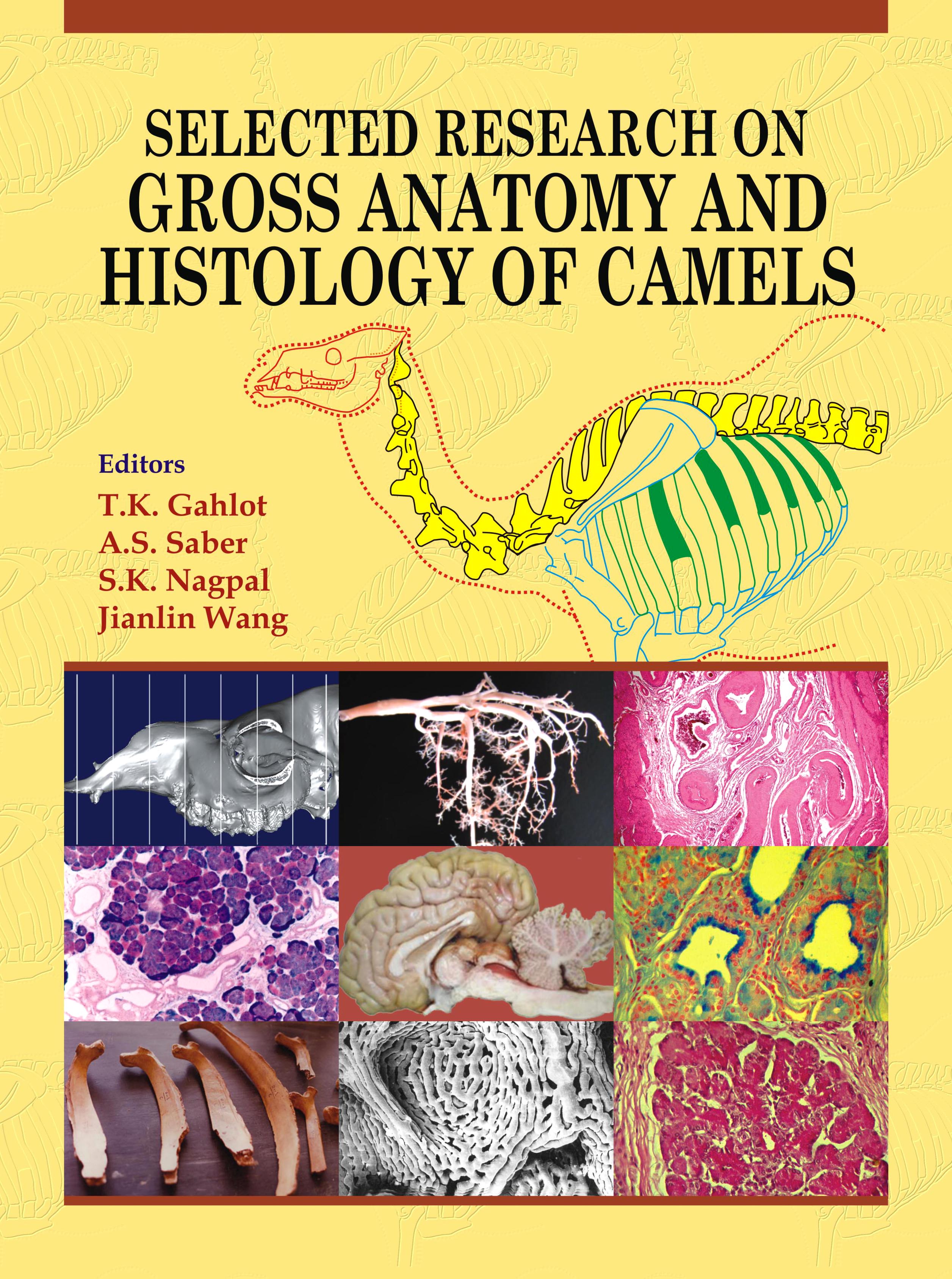
Selected Research on Gross Anatomy and Histology of Camels is a dream come true for its editors and the book comprises 92 manuscripts spread over 9 sections. The manuscripts related to dromedaries and bactrians were 75% and 25%, respectively. Major quantum of work was done on digestive system (17.39%), followed by radiological anatomy (16.3%), circulatory system (14.13%), respiratory system (14.13%), urogenital and nervous system (10.86%, each), and skeletal system and common integument (4.35%, each). Miscellaneous papers (7.61%) made a separate group. Bactrian camel research remained focused mainly on respiratory, urogenital and nervous system.
This book offers an advantage of exclusive histology of several organs (30.43%) and scanning electron-microscopy (14.13%) in few manuscripts. Special staining depicted in few histomorphological pictures adds to wide spectrum of histological details of many organs. Radiographic anatomy was studied through radiography (plain and contrast) and magnetic resonance imaging (MRI). More emphasis was given on skeletal system followed by circulatory, nervous, dentition (teeth) and digestive system. Anatomy of skeletal system was studied through morphological features only. Digestive system was studied by gross, histological, scanning and transmission electron microscopy and histochemical methods.
The histology, histochemical and immunohistochemical based manuscripts have a wide range of photomicrographs using a variety of special stains and methods. These were Gordon and Sweet, H & E, PAS-AB, Ojawa and Mayahara, Modified tetrazolium, Gomori’s Aldehyde Fuchsin, Phosphotungstic acid haematoxylin, Hellerstrom and Hellman’s silver, Mayer’s Mucicarmine stain, Toluidine blue stain, Van Gieson, Verhoeff, Weigert’s, Mallory’s Trichrome, Hoechst 33342, McManus’s, Gomori Acid Phosphatase, Zinc Iodate Osmium Tetroxide and Bielschowsky.
Anatomy of respiratory system was perhaps studied more in bactrian camels than dromedaries. The parts studied were larynx, nasal cavity, paranasal sinuses, caudal choanae, trachea and pulmonary tissue. Anatomy of urogenital system section included gross and histological study of various male and female genital organs, kidneys, glomerulus, etc.
Anatomy of circulatory system was well studied by researchers. Arterial and venous supply of various organs was documented. Erythrocyte morphology and ultrastructure of formed elements of blood were studied in detail. Research on lymphoid tissue and microvascularization of placenta were also a hallmark of research.
Anatomical details of brain, spinal cord and spinal nerves of dromedary and bactrian camels are important highlights of this section on nervous system. Morphometric and microscopic evaluation of heart, kidneys and adrenal glands of camel calves, adipocyte pattern of camel hump and kidneys, cytoenzymic studies on the blood cells in dromedary camels and functional anatomy of external and middle ear of bactrian camels marked the miscellaneous section of this unique book.
Editors view this book as a milestone in the field of camel anatomy, as it provides histology, special staining and electron-microscopy and advanced imaging too of few organs or systems of dromedary and bactrian camels. We hope that this book proves an important reference book for all future researchers in the field of camel anatomy.
Camel Publishing House is thankful to Dr.KapilKachwaha, Dr. Anita Meena and
Sh. Kheraj Ram Jat for their skillful editorial assistance in shaping the manuscripts of this book.
T.K. Gahlot
A.S. Saber
S.K. Nagpal
Jianlin Wang
(Editors)
 Home
Home