Preface
The Journal of Camel Practice and Research (JCPR) started publishing in 1994 as a logical response to the rising interest in the camelids. It was hailed as a significant development in the camelid world. In its 15 years of biannual publication since then it has become the largest single resource of camelid literature. Despite this, there had been a persistent paucity of subject specific information compiled in the form of a book or compendium. It was precisely to fulfil this need that the Camel Publishing House has taken the initiative to make available such specialised resource material to camelid practitioners and researchers. As a result, the first such compilation was published in 2004 under the title, ‘Selected Research on Camelid Physiology and Nutrition’. This book received a very good response as it must have updated the knowledge of scientists as well as facilitated researchers with valuable information in a classified from. Additionally it helped identifying the world wide laboratories/institutions and personnels engaged in research of those particular field.
Scanning of papers published in JCPR in last 15 years revealed that articles related to parasitology were 11.33% and it emerged as the 3rd major subject of research during these years. To date, there is no exclusive book on camelid parasitology. Camelid parasitology has made new strides in recent years which are evident by introduction of newer diagnostic techniques and treatments. Researchers have also studied the immunological aspects of parasitic diseases. There are at least 17 countries involved in camelid parasitology research, viz. Ethiopia, France, India, Iran, Jordan, Kenya, Libya, Mauritania, Nigeria, Sultanate of Oman, Pakistan, Saudi Arabia, Sudan, Sweden, United Arab Emirates, Uganda and U.S.A. As per published papers in JCPR, 173 authors have contributed these manuscripts. Out of these Kenya ranks number one (34 authors) followed by India (32), Pakistan (17), USA (16), Iran (15), Nigeria (6) and Saudi Arabia (10) in order. The country-wise laboratories/institutions involved in camelid parasitology research are given in appendix 1 and names of scientists involved in appendix 2. Kenya Trypanosomiasis Research Institute (KETRI), P.O. Box 362, Kikuyu, Kenya has emerged as biggest resource of camelid parasitology research during this period. In India, Department of Veterinary Parasitology, College of Veterinary and Animal Science, Bikaner-334001, Rajasthan has emerged as biggest contributor of camelid parasitology research followed by Department of Veterinary Parasitology, Haryana Agricultural University, Hisar (Haryana), India.
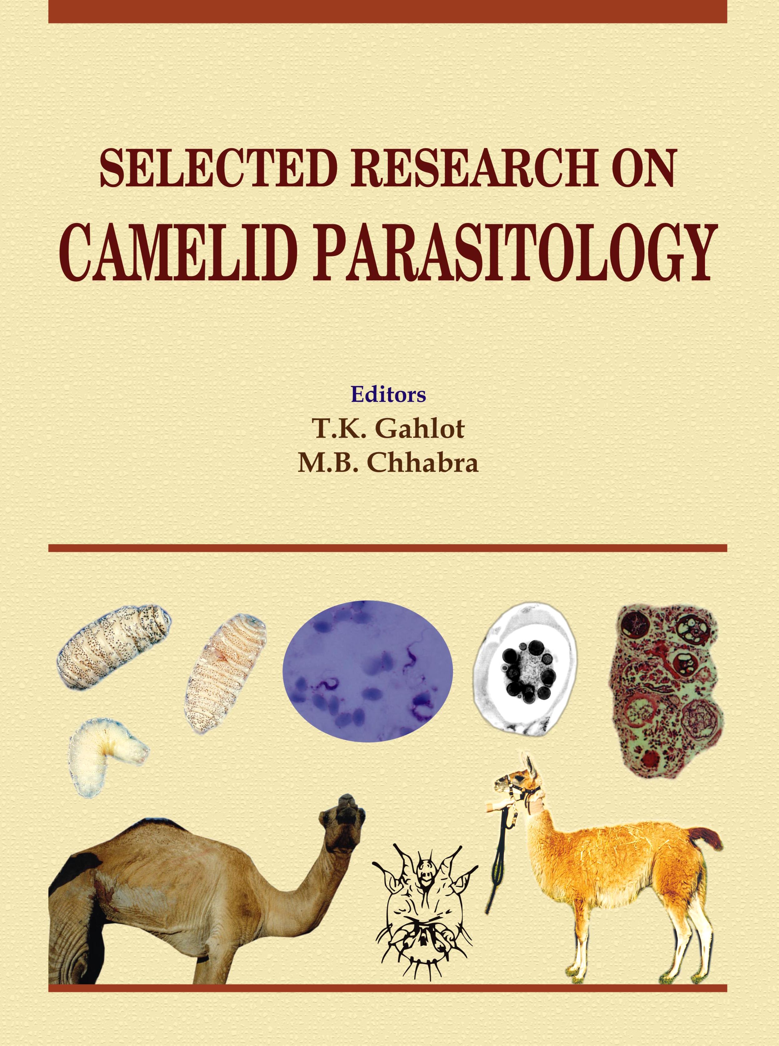
An overview of contents of this book (appendix 3) suggests that global camelid parasitology research was more focussed on protozoa (39%), followed by arthropods (24%) and helminths (18%). Among the protozoa, maximum research remained focussed on trypanosomiasis (31%) and among arthropods maximum thrust had been on mange (12.5%). New World camelids found only 10% share of camelid parasitology research while remaining 90% were related to dromedary and none to bactrians. Notable contributions were a 3-part review series authored by Prof.M.B.Chhabra and his colleagues on parasites and parasitic diseases (Protozoan, Helminthic and Arthropods) of dromedary camels.
Trypanosome infection was diagnosed by various diagnostic tests including parasitological, chemical, biological and immunodiagnostic tests. Comparison of various tests included MIT followed by ag-ELISA, WFE, MCT, TFE, and AGIDT. The usefulness of combining Suratex® and parasitological tests for pen-side diagnosis and chemotherapeutic control of surra was also studied. Antigen-detection ELISA was positive in all the parasitologically proven cases as well as those with no parasitaemia and was found to be more useful method for the diagnosis of latent surra in camels. Sero-immunological studies revealed a significant reduction (p<0.05) in haemolytic complement and an increase in complement fixing antibodies in infected cases of trypanosoma. Complement fixing antibody titres and circulating trypanosomal antigen levels were also monitored throughout the infection period. A rapid initial increase (47%) in mean alternative pathway haemolytic complement (ACH50) level was seen during the first week of infection. The third complement component (C3) was isolated from camel serum by polyethylene glycol precipitation and chromatography on DEAE-Sephadex A-50, CM-Sephadex C-50 and Sephacryl S-200. The hypocomplementaemia in T. evansi infected camels was attributed to the presence of the parasite. It was opined that camel C3 is a high molecular weight protein and that its depletion (activation) occurs in trypanosome infected camels.
Drug resistance and treatment of Trypanosomaevansi against quinapyramine sulphate, Triquin® (Quinapyramine sulphate/chloride-Prosalt) and Cymelarsan were studied. Diminazeneaceturate formulation, Trypan® was found effective in treatment of camel trypanosomiasis. Drug resistance to older trypanocides such as Suramin and Trypacide, and the efficacy of new ones such as Melarsomine were studied, with a view to provide epidemiological data and advice on the most appropriate drug regimes for management of the disease in camels. Trypanosomiasis was extensively studied in Mauritania, Iran, India, Kenya, Pakistan and Eastern Africa.
Gastrointestinal parasites were detected in faecal samples by direct method examination, floatation method and sedimentation method. Egg output was quantified by means of the Mc Master technique. Nine types of parasites were diagnosed. Trichostrongylusprobolurus, Camelostrongylusmentulatus and Cooperiaonchophora were the most common. Fasciola hepatica was recorded for the first time in Jordanian camels. Nematode and cestode infections in slaughtered camels in Pakistan were 34.66 and 8%, respectively. The nematodes found were Haemonchuslongistipes(12.5%), H. contortus(6%), Trichurisovis(2%), T. globulosa(1.5%) and Oesophagostomumvenulosum(2%). Cestode infection recorded were Monieziaexpansa(3.33%) and M.benedeni(2%). A haemato-biochemical study was also conducted in camels harbouring natural mixed infection of gastrointestinal (GI) nematodes (Haemonchuslongistipes, Trichostrongylusspp,Strongyloidespapillosus, NematodirellasppandTrichurisspp)with epg of 1400-28000. Comparative efficacy of anthelmintics against mixed infection of Haemonchuslongistipes, Trichostrongylusspp, Strongyloidespapillosus, Nematodirellaspp and Trichurissppin camel were evaluated. Ivermectin showed highest efficacy (98.96 99.43%) followed by albendazole (98.95 - 99.30%), levamisole (98.03 -98.38%), fenbendazole (94.23 - 99.65%) and tetramisole (96.40 - 98.50%). Albendazole and fenbendazole did not show useful effect against Trichuris. Another study indicated that fenbendazole and morantel citrate at the dose rate of 5.0 mg/kg body weight have 98 and 91.01% efficacy, and at 7.5 mg/kg body weight have 100 and 92.79% efficacy, respectively against mixed gastrointestinal nematodoses in camels of Rajasthan. Efficacy of ivermectin (injectable) and levamisole hydrochloride (injectable) against natural strongyle nematode infection in dromedary camels was evaluated over a period of three months and a reduced efficacy of ivermectin to treat strongyle nematodes was found which could be indicative of the development of resistance by these worms to the drug.
Prevalence of Dipetalonemaevansi in four herds of camels situated in different villages of Bikaner district and prevalence and pathogenic effect caused by O.fasciata during tissue invasion phase in camels in Islamic Republic of Iran has also been reported.
The section of arthropods and allied contains majority of papers related to mange. Prevalence of sarcoptic mange in camels in Borno state of Nigeria, camel mange affecting humans, experimental mange, and histopathology of mange affected camel skin formed part of some manuscripts. Immunological studies included use of radioimmunoassay (RIA) methods and significantly higher levels of IgE were recorded in camels suffering from mange. Specific antibodies to S.scabieiwere confirmed in a western blot analysis. The S. scabieivarvulpesantigen employed in the ELISA was analysed by polyacrylamide gel electrophoresis (PAGE) in the presence of sodium dodecyl sulphate (SDS) under reducing conditions and by western blot (WB) using a Mini-Protean system.
The efficacy of various acaricidal drugs such as Amitraz 12.5%, Sebacil E.C.50%, Gamatox and Ivomec in the treatment of advanced and well established lesions of sarcoptic mange in the one-humped camel was evaluated.
Prevalence of nasal myiasis by the larvae of the camel nasal bot fly, Cephalopinatitillator, tsetse transmitted trypanosomosis, infection with Linguatulaserrata nymphs and occurrence of Limnatisniloticain camels made part of some other manuscripts of this section.
An interesting part of this book is a section on parasitism of New World camelids. Lamanemachavezi is a trichostrongyle that differs from others of the family in that the larvae migrate to the liver and lungs as part of the life cycle. Parasitism is serious in young Alpacas in Peru. Other trichostrongyle nematodes currently found only in South America include Graphinemaaucheniaeand Spiculopteragiaperuviams. Aberrant parasites of SACs in North America may cause serious regional problems.Parelpahostrongylustenuis (meningeal worm) is a relatively non-pathogenic parasite of the white tailed deer Odocoileusvirginiams, which is present throughout much of the eastern third of the USA. In the SACs migration of the larvae through the spinal cord produces paresis and paralysis. In SACs, the larvae ofCephenemyaspp (deer nasal bot fly) cause the development of a granuloma in the nasopharynx that may obstruct airflow. An experiment demonstrated that a camel T. evansi strain was able to infect a guanaco, inducing typical signs of subacuteSurra. Gastrointestinal nematodes, meningeal worm (Parelaphostrongylustenuis), Lamanemachavezi, Nematodirusspp and whipworms (Trichurisspp) infected SACs. Three llamas with suspected cerebrospinal nematodiasis caused by aberrant larval migration of Parelaphostrongylustenuis were treated with moxidectin. Prevalence of parasites in llamas kept at Surman Park in Libya showed no external parasites and haemoparasites. Faecal examination revealed Nematodirusspp, Trichostrongyle and Strongyloidesspp, Trichurisspp, unidentified large egg, Fasciolahepatica and Eimeria spp. Another study on ten llamas naturally infected with gastrointestinal parasites, e.g. Nematodirus, Capillaria and Strongyloides species were treated with albendazole. An eleven year old male alpaca (Lama pacos) showing episodes of seizure-like activity revealed numerous nymphal and adult ticks in the left external ear canal adjacent to the tympanic membrane. Those were identified as Otobiusmegniniongross necropsy.
The current volume entitled as “Selected Research on Camelid Parasitology” will prove a milestone compilation of diverse manuscripts on new and old world camelids. This would go a long way in helping researchers to update information base in their area of study. We are sure that future researchers would make an endeavour by updating the three review papers mentioned in this volume after every 5 years. We thank all the contributors of manuscripts for enriching the contents of this valuable book.
Dr. T.K. Gahlot
Dr. M.B. Chhabra
(Editors)
 Home
Home